
Keratoconus is a progressive eye condition that affects the shape and structure of the cornea, leading to significant problems with vision. As the disease advances, the cornea becomes steeper, causing vision to decline further.
We offer a groundbreaking treatment for keratoconus, known as cross linking eye surgery. This innovative procedure strengthens the cornea naturally, halting the progression of the condition without requiring invasive eye surgery.
By utilising the natural interaction between riboflavin and ultraviolet light, cross linking eye surgery creates new bonds between collagen fibres in the cornea, helping to preserve vision. For those concerned about keratoconus, this treatment provides a scientifically proven method to stabilise the cornea and potentially prevent further deterioration of vision. To find out more about the pricing details, please contact us for more information.
Key Takeaways
- Cross linking eye surgery is a groundbreaking treatment for keratoconus.
- This procedure strengthens the cornea naturally, halting disease progression.
- It provides a minimally invasive option to preserve vision.
- The treatment utilises riboflavin and ultraviolet light to create new collagen bonds.
- Patients can benefit from a scientifically proven method to stabilise the cornea.
Understanding Keratoconus and Its Impact on Vision
Keratoconus, a progressive eye disease, requires prompt attention to preserve vision. It is a condition that affects the cornea, the clear dome-shaped surface at the front of the eye, causing it to thin and bulge. This alteration in the cornea’s shape can significantly impact vision, leading to distortion and blurriness.
What is Keratoconus?
Keratoconus is characterized by the cornea taking on a cone-like shape, rather than its normal rounded shape. This irregular curvature of the cornea deflects light as it enters the eye, resulting in distorted vision. “The cornea plays a crucial role in focusing light, and any irregularity in its shape can lead to significant visual disturbances,” as noted by eye care professionals.
The progression of keratoconus can vary among patients, with some experiencing rapid deterioration in vision, while others may have a slower progression over many years.
Who is Affected by Keratoconus?
Keratoconus typically affects young people, with diagnosis most commonly occurring in the late teenage years or early twenties. Research indicates that certain demographic groups, particularly those of Black and Asian heritage, have higher rates of keratoconus. There appears to be a genetic component, with approximately 10% of patients reporting a family history of the condition. In the UK, approximately 1 in 2,000 people are affected by keratoconus, resulting in varying degrees of severity from mild cases requiring only glasses to advanced cases that may impact daily functioning.
Both men and women can develop keratoconus, though some studies suggest a slightly higher prevalence in male patients. Certain factors may increase the risk, including chronic eye rubbing, allergic conditions affecting the eyes, and connective tissue disorders.
What is Cross Linking Eye Surgery?
For individuals suffering from keratoconus, cross-linking eye surgery offers a beacon of hope by strengthening the cornea. This minimally invasive procedure has revolutionised the treatment of keratoconus, a condition where the cornea progressively thins and bulges, leading to distorted vision.
Cross-linking eye surgery involves the use of riboflavin (vitamin B2) and ultraviolet (UV) light to create new bonds between the collagen fibres within the cornea. This process enhances the cornea’s rigidity and stability, halting the progression of keratoconus.
The Science Behind Corneal Cross-Linking
The science behind corneal cross-linking is rooted in the interaction between riboflavin and UVA light. When the riboflavin-soaked cornea is exposed to UVA light, it triggers a chemical reaction that forms new bonds between the collagen fibres. This reaction strengthens the cornea, making it more resilient to the distortions caused by keratoconus.
The ultraviolet light and riboflavin react together to create cross-links within the cornea, which make it stiffer, mimicking the normal age-related cross-linking effect that occurs from your 30s onwards.
How Cross-Linking Strengthens the Cornea
Cross-linking works by creating new bonds between collagen fibres in the cornea, effectively reinforcing its internal structure much like adding extra support beams to a weakening framework. The procedure increases the cornea’s rigidity by approximately 300%, helping it to maintain its proper shape and resist the progressive bulging characteristic of keratoconus.
- During cross-linking, the riboflavin-soaked cornea is exposed to precisely calibrated UVA light, which activates the riboflavin molecules to form new chemical bonds between adjacent collagen strands.
- These newly formed cross-links act as “anchors” that strengthen the corneal tissue, preventing further thinning and distortion without changing the cornea’s transparency.
- Unlike corneal transplants that replace tissue, cross-linking preserves the patient’s own cornea by enhancing its natural structure, making it a much less invasive approach to treating keratoconus.
The strengthening effect of cross-linking typically stabilises within 3-6 months after the procedure, though the full remodelling process of the cornea may continue for up to a year. By understanding how cross-linking works, patients can appreciate the innovative approach it offers in managing keratoconus.
When is Cross Linking Eye Surgery Recommended?
Identifying the optimal time for cross-linking eye surgery involves careful monitoring of keratoconus progression. We use a combination of corneal scans, vision tests, and spectacle prescription to look for changes in a patient’s condition. If we detect a change, we can typically list the patient for the procedure within a few weeks.
Early Signs That Indicate Treatment Need
Several early signs may indicate the need for cross-linking eye surgery. These include noticeable changes in vision, such as increased astigmatism, and significant progression of keratoconus. Prompt intervention can help prevent further deterioration.
Diagnostic Tests for Keratoconus Progression
Various diagnostic tests are employed to monitor keratoconus progression. These include:
- Corneal topography, which provides detailed maps of the cornea’s surface, detecting subtle changes in shape and thickness.
- Sequential corneal imaging, allowing precise measurement of progression by comparing scans taken months apart.
- Changes in corneal thickness measurements (pachymetry), which are carefully monitored as progressive thinning is a key indicator of worsening keratoconus.
- Regular refraction tests tracking changes in spectacle prescription, particularly increasing astigmatism.
- Visual acuity measurements with both glasses and specialised contact lenses to assess functional vision changes.
For patients showing documented progression on these diagnostic tests, we typically recommend scheduling cross-linking treatment promptly to prevent further deterioration.
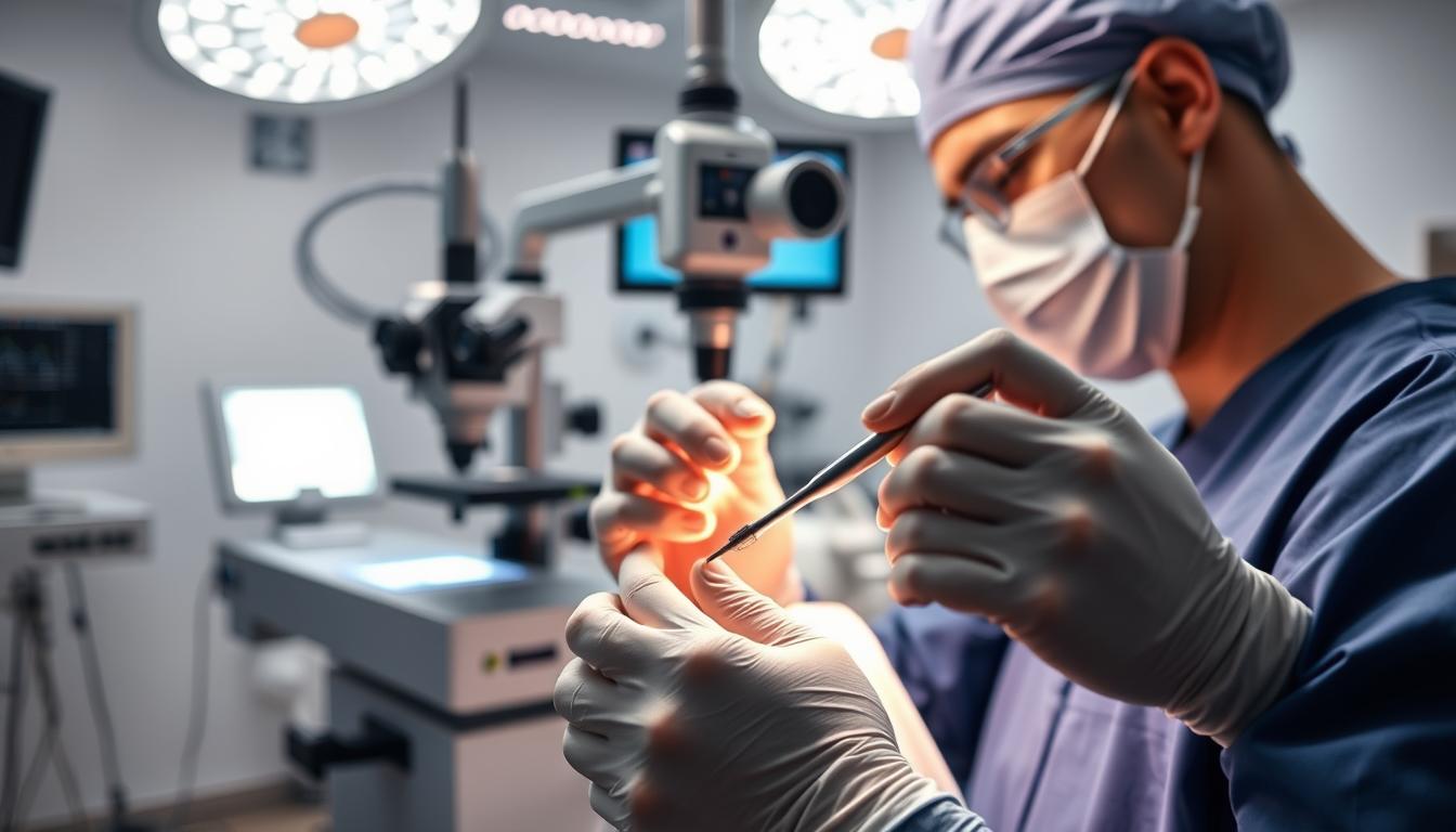
The Cross Linking Eye Surgery Procedure Explained

Understanding the cross linking eye surgery procedure is crucial for patients considering this treatment option. The procedure involves a series of steps that work together to strengthen the cornea and halt the progression of keratoconus.
Step-by-Step Process
The cross linking eye surgery procedure is typically performed in a clinical setting. The process begins with the application of anaesthetic eye drops to numb the eye surface, ensuring the patient’s comfort during the treatment. The medical team then proceeds to apply a riboflavin solution to the cornea, which is subsequently exposed to UV light. This exposure triggers a cross-linking reaction that strengthens the corneal tissue.
Duration and What to Expect During Treatment
The entire cross linking treatment typically takes about 60-90 minutes per eye, although the actual UV light exposure lasts only 8-10 minutes. During this time, patients are asked to lie still and focus on a fixation light. The medical team provides reassurance and guidance throughout the process, ensuring the patient’s comfort and safety.
At the conclusion of the procedure, a bandage contact lens is placed on the eye to protect the treated area and enhance comfort during the initial healing phase. Patients can expect to go home the same day after receiving detailed aftercare instructions.
Benefits of Cross Linking Eye Surgery
Cross-linking eye surgery offers numerous benefits for patients suffering from keratoconus. By strengthening the cornea, this treatment can significantly improve the quality of life for those affected.
Preventing Disease Progression
The primary goal of cross-linking eye surgery is to stabilise the cornea and prevent further deterioration of vision. This is achieved by creating new bonds within the corneal tissue, effectively halting the progression of keratoconus. As a result, patients can avoid more severe vision problems in the future.
Potential Vision Improvements
While the main objective is to stop disease progression, many patients experience improved vision as a secondary benefit. Approximately 50-60% of individuals report some degree of vision improvement following treatment, though this varies between individuals. Improvements in the shape of the cornea can lead to better visual acuity, and in some cases, a reduction in refractive error. Overall, the results can be quite positive, making cross-linking a valuable treatment option.
- Vision improvements typically occur gradually over 6-12 months.
- Some patients experience a reduction in their refractive error.
- The flattening effect of cross-linking can reduce irregular astigmatism.
Recovery After Cross Linking Eye Surgery
Understanding the recovery process after cross-linking eye surgery can help manage expectations and lead to better results. The recovery journey is just as important as the surgery itself, involving several stages that are crucial for achieving optimal vision.
Immediate Post-Procedure Care
Immediately after the surgery, your eyes will be sensitive, and you may experience discomfort. It’s essential to follow the post-procedure instructions provided by your doctor to ensure proper healing. This typically includes using medicated eye drops and wearing a bandage contact lens to protect the eyes.
First Week of Recovery
During the first week, your vision may be blurry, and you might experience some discomfort. A follow-up appointment is usually scheduled within the first week to remove the bandage contact lens and assess the healing progress. As one patient reported, “I could see about 80% of what my original vision was after the bandage lenses were removed.”
Long-Term Healing Process
The complete healing process extends over several months, typically between 6 to 12 months, as the cornea continues to remodel. Most patients can return to wearing glasses within 1-2 weeks, though the prescription may need adjustment as the cornea stabilises over the following months. Vision typically improves gradually over 3-6 months, with some patients continuing to notice improvements for up to a year after the procedure.
Potential Risks and Side Effects
While cross-linking eye surgery is generally considered safe, it’s crucial to understand the potential risks and side effects associated with this treatment. As with any surgical procedure, there are complications that can arise, and being informed is key to managing expectations and outcomes.
Common Temporary Side Effects
After undergoing cross-linking eye surgery, patients may experience some temporary side effects. These can include discomfort, sensitivity to light, and blurred vision. Most of these issues resolve on their own within a short period. However, some patients may experience persistent corneal haze, which occurs in approximately 2-3% of cases and may require additional treatment if it significantly affects vision.
It’s also worth noting that delayed epithelial healing can increase the risk of infection and scarring, particularly in patients with pre-existing conditions like dry eye syndrome. We take every precaution to minimize these risks through careful patient screening and post-operative care.
Rare but Serious Complications
Although rare, there are serious complications that can occur with cross-linking eye surgery. Corneal infection (keratitis) is a potential serious complication that requires prompt treatment with intensive antibiotic therapy to prevent permanent scarring. In rare instances, approximately 3% of cases, patients may experience a decrease in vision directly related to the cross-linking procedure, often due to corneal scarring or irregular healing.
In extremely rare cases, damage to the corneal endothelium can occur if safety protocols regarding corneal thickness are not strictly followed. We adhere to stringent safety protocols to minimize these risks and provide close follow-up care to address any complications promptly.
It’s essential to note that there’s about a 3% risk of someone’s vision being worse directly as a result of the cross-linking treatment. However, with proper care and follow-up, the majority of patients undergo the procedure without experiencing significant complications.
Patient Experiences with Cross Linking Eye Surgery
As we explore patient experiences with cross-linking eye surgery, it becomes clear that this procedure offers more than just a medical solution. Patients’ journeys through cross-linking eye surgery are as unique as they are, yet common themes emerge that highlight the procedure’s impact on their lives.
The Procedure from a Patient’s Perspective
Patients often describe their experience of undergoing cross-linking eye surgery as a positive one, with many expressing relief at having taken proactive steps to address their keratoconus. As one patient noted, “First impressions are yes [I am happy with how it went] because I’m healing every day, and I feel like it’s going the way it should have done.” This sentiment is echoed by many, who appreciate the procedure’s potential to stabilise their vision and prevent further deterioration.
Recovery Journey and Results
The recovery journey following cross-linking eye surgery is typically described as gradual, with comfort levels improving significantly within the first few days. However, it may take longer for vision to stabilise. Key aspects of the recovery process include:
- Returning to desk work or light activities within 3-5 days, with precautions such as wearing sunglasses and using lubricating eye drops.
- Experiencing improvements in vision stability over time, which is frequently cited as the most important outcome.
- Noticing actual improvements in vision over the months following cross-linking, although this is not guaranteed.
Many patients become advocates for early intervention, recommending cross-linking eye surgery to others with keratoconus based on their positive experiences. The peace of mind that comes from halting disease progression is a significant psychological benefit, reducing anxiety about future vision loss. As patients reflect on their journey, they often express gratitude for the procedure’s impact on their vision and overall quality of life.
Conclusion: Taking the Next Step Towards Preserving Your Vision
Ultimately, cross linking eye surgery emerges as a crucial treatment for keratoconus, offering patients a pathway to stabilised vision. Early intervention is key to achieving the best possible outcomes, making timely diagnosis and treatment essential. The minimally invasive nature of this procedure makes it an attractive option. While there is some temporary discomfort and time needed for recovery, the long-term benefits make it worthwhile. For personalised advice, we encourage you to contact our team to arrange a comprehensive consultation. During this consultation, we can discuss your specific case and provide information about treatment timing and expectations. To learn more, please contact our patient care team directly.
FAQ
What is the purpose of using a bandage contact lens after corneal cross-linking treatment?
We use a bandage contact lens to protect the cornea and promote healing after the procedure. It helps to reduce discomfort and pain, and is usually removed within a few days.
How long does the corneal cross-linking procedure take?
The treatment typically takes about 30 to 90 minutes per cornea. The duration may vary depending on the individual case and the specific technique used.
What is the role of riboflavin in corneal cross-linking?
We apply riboflavin drops to the cornea, which are then activated by a specific wavelength of light. This process strengthens the collagen bonds within the cornea, helping to halt the progression of keratoconus.
Will I experience pain after the procedure?
We may prescribe pain management medication to help alleviate any discomfort. Some patients may experience mild pain or sensitivity, but this typically subsides within a few days.
How long does it take to recover from corneal cross-linking?
The initial healing process usually takes a few days to a week. However, it may take several months for the cornea to fully stabilise and for vision to improve.
Can corneal cross-linking prevent the need for a corneal transplant?
In many cases, corneal cross-linking can halt the progression of keratoconus and potentially avoid the need for a corneal transplant. However, this depends on the individual case and the severity of the condition.
Are there any potential risks or complications associated with corneal cross-linking?
As with any medical procedure, there are potential risks, including infection, scarring, and temporary vision disturbances. We will discuss these risks in detail during your consultation.
How effective is corneal cross-linking in treating keratoconus?
Corneal cross-linking has been shown to be highly effective in halting the progression of keratoconus and improving vision in many patients. Results may vary depending on the individual case.

